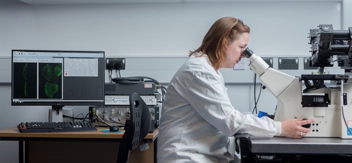
Light sheet microscopy
Overview
Light sheet Fluorescence Microscopy (LSFM) or Selective Plane Illumination Microscopy (SPIM) is a rapidly developing imaging technique that allows for increased imaging speeds with little or no photo-bleaching and photo-toxicity. The technique features a unique lens configuration where the excitation lens is positioned perpendicular to the detection lens, thereby confining the excitation of the sample only to the volume being observed. Consequently, LSFM is perfectly suited to 4D imaging of live single cells and smaller organisms. Conventional imaging methods such as widefield and confocal microscopies use an illumination configuration that exposes the entire sample to illuminating radiation. In live cell imaging, this whole cell illumination strategy can be detrimental to the physiological state of the specimen.
Access to the COMPARE tissue culture suite for preparation of living samples is available.

In COMPARE at the University of Birmingham Imaging Suite we have three advanced LSFM systems; Lattice Light Sheet Microscope, Marianas diSPIM microscope and the Ultramicroscope ll.